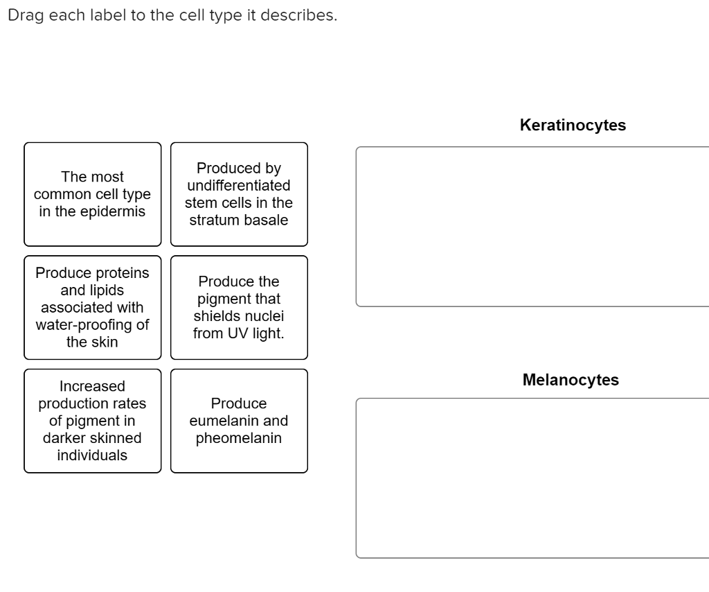
Drag Each Label to the Cell Type It Describes.
Include descriptions of what each organelle does. Describe some of the characteristics of living things.

Vince Camuto Thanley Pointed Toe Pump Nordstrom In 2022 Pumps Pointed Toe Pumps Heels
Drag each label to the appropriate target.
. Create a cell diagram with each part of plant and animal cells labeled. Drag each label to the cell type it describes Keratinocytes The most common cell type in the epidermis Produced by undifferentiated stem cells in the stratum basale Produce proteinsProduce the and lipids associated. Uses energy growth and development made of cells eat reproduce.
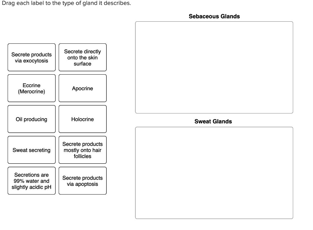
The first step in preparing for division is to replicate the cells DNA in the S phase. Same thing that was listed above. Drag each label to the type of gland it describes.
Never have a cell wallhave chloroplastsare prokaryoticsometimes have a cell wallhave a nucleoidalways have a cell wall. Up to 24 cash back List the organelles and approximate size of the cells in each sample. Holocrine Sebaceous gland or Sudoriferous Glands.
Similarly any wastes produced within a prokaryotic. Most mammalian red blood cells have no nucleus. Describe how the skin cells neurons muscle cells and blood cells you have observed.
Gizmo Warm-up In the Cell Types Gizmo you will use a light microscope to compare and contrast different. T cells Capable of developing Differentiate into memory cells plasma cells when activated Mature in thymus Lymphocytes Provide a defense B cells against extracellular Arise originally from antigens rather than bone marrow directly attack them Attack the foreign The. Drag the labels to the appropriate bins to identify the step in protein synthesis where each type of RNA first plays a role.
Drag each label to the type of gland it describes. Some cells are only triggered to enter G 1 when the organism needs to increase that particular cell type. Once a cell passes the G1 checkpoint it usually completes the cell cyclethat is it divides.
Humans plants and mushrooms are all alive. A tissue is a group of similar cells that together carry out a specific function. Each depression has a label of A B or RhD.
One tray is used for each blood sample. The cell plate grows toward and eventually fuses with the cell wall of the parent cell5. At 0150 µm in diameter prokaryotic cells are significantly smaller than eukaryotic cells which have diameters ranging from 10100 µm Figure 37.
Compares the genomes of different organisms compares the limbs of different organisms compares fossilized structures to living organisms compares cells of organism Comparative Anatomy Molecular Biology 2. Drag each label to the. For example anti-A antiserum containing anti-A antibodies goes into the depression.
Neutrophils are the most common type of white blood cell in the body with levels of between 2000 to 7500 cells per mm 3 in the bloodstream. For each blood sample. These cells exit the cell cycle before the G1 checkpoint.
Using arrows and Textables label each part of the cell and describe its function. Many types of cells such as the ones in this activity li ve together in groups called tissues. Drag each label into the appropriate position to identify what cell type is described by the label.
Place a drop of blood in each of the three depressions of one testing tray. Find diagrams of a plant and an animal cell in the Science tab. The majority of the cells arise as keratinocytes from stem cells in the stratum basale and migrate outwards as new cells are regenerated to replace the sloughed exfoliated cells of the.
Many organisms contain cells that do not normally divide. Question 9 Each label describes one or more of three types of connective tissue cells. Zonula occludens describes that there is a formed band of tight junctions which encircles every cell.
Sort the cell features based on the type of organism they describe. Drag each label to the correct location on the chart. These types are mostly present in a given order at apical ends of epithelial cells Tight or occluding junctions edit edit source This type of junction is also called zonula occludens and is the most apical structure in the epithelial cell.
Color the text boxes to group them into organelles found in only animal cells organelles found. Many cells temporarily enter G 0 until they reach maturity. The bones are a core founding component of a living body that holds the structure of muscles and organsThe bones of the skeletal system are composed of two types of tissues ie compact and spongy bone tissue.
A tissue is a group of similar cells that together carry out a specific function. If an RNA does not play a role in protein synthesis drag it to the not used in protein synthesis bin. Drag each label to the correct location.
Below detailed information about each type will be discussed. What do these organisms have in common. Describe the two fields of study.
The small size of prokaryotes allows ions and organic molecules that enter them to quickly spread to other parts of the cell. Match the function to the type of tissue. Place a drop of the antiserum that is associated with each depression.
Find an answer to your question Drag each label to the correct location on the chart. The Compact bone tissue covers the outer part of the bone structure and provides toughness and strength to the structure of bone. This allows the red blood cell to use all of its volume to transport oxygen.
Types of Bone Cells. Extend your thinking. TranscriptionRNA processing- pre-mRNA mRNA snRNA translation-.
Neutrophils are medium-sized white blood cells with irregular nuclei and many granules that perform various functions within the cell. Many types of cells such as the ones in this activity live together in groups called.

It S Real It S Called For Want Of A Nail It S On Ao3 Merlin Show Merlin Fandom Merlin

Solved Drag Each Label To The Cell Type It Describes 3 34 Chegg Com

Difference Between Phagocytosis And Pinocytosis Teaching Biology Biology Classroom Cell Biology Notes

Solved Drag Each Label To The Type Of Gland It Describes Chegg Com
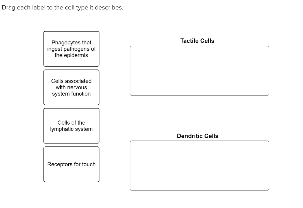
Solved Drag Each Label To The Cell Type It Describes Chegg Com

Water Cycle Diagram Water Cycle Diagram Cycle Drawing Water Cycle

Ahcdweek2sol10 Pdf 10 Award 10 00 Points Problems Adjust Credit For All Students Drag Each Label To The Cell Type It Course Hero

Pin By Manuel Garcia On Virologia Viral Infection Medical Nursing Students

Mitosis Drag And Drop Mitosis Teaching Cells Mitosis Activity
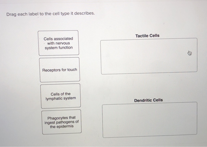
Solved Drag Each Label To The Cell Type It Describes Chegg Com

Easy Method For Making A Photosynthesis Concept Map With Example Photosynthesis Photosynthesis Activities Concept Map
![]()
Edcite S Essay Response Question Type Essay No Response Language

Chapter 6 Worksheet Flashcards Quizlet

Edcite S Essay Response Question Type Essay No Response Language
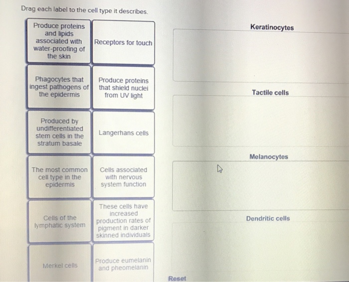
Solved Drag Each Label To The Cell Type It Describes Chegg Com

Microsoft Announces Patches For 3 Critical Windows 8 1 Flaws Ignores Office Zero Day Vulnerability The Tech Journal Sharepoint Microsoft Office Microsoft

Module 2 Chapter 6 Flashcards Quizlet

Chapter 6 Worksheet Flashcards Quizlet
Solved Drag Each Label To The Cell Type It Describes Chegg Com
Comments
Post a Comment